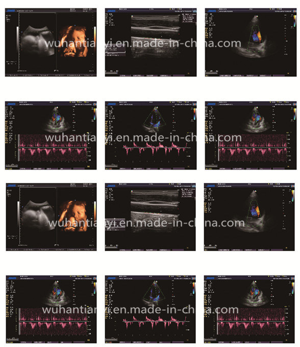
An excellent HCU (Hand-Carried Ultrasound) System TY-6868D is a High-end Color Doppler HCU (Hand-Carried Ultrasound). Based on Tianyi's leading digital ultrasound technology, TY-6868D offers superb 2D and premier doppler imaging modes such as CFM, CW, PW, CDE, CCD, HPRF.
Features & Advantages:
Multi-application: abdomen, cardiology, OB/GYN, small parts, peripheral vessels, urology, rectum, pediatric, orthopedic, intra-operative ultrasound, ultrasound guided biopsy.
Continuous high-precision DBF
THI (Tissue Harmonic Imaging)
TDI (Tissue Doppler Imaging)
Panoramic zoom
Parallel scanning
PC platform / Windows XP Embedded O/S
Standard Configuration:
TY-6868D main unit 15" high definition non-interlaced LCD monitor, special for medical imaging DICOM 3.0 Measurement & calculation software packages
Electronic convex array transducer: CA3.5MHz/R50 (2.0-6.0MHz)
Electronic linear array transducer: LA7.5MHz/L40 (5.0-10.0MHz)
Options:
Electronic linear array transducer: LA7.5MHz/L50 (5.0-10.0MHz)
Phased array transducer: PA2.5MHz (2.0-4.0MHz)
Electronic endocavity transducer: EV6.5MHz/R10 (5.0-9.0MHz)
Electronic micro-convex array transducer: MC3.5MHz/R20 (2.0-6.0MHz)
Needle-guided brackets
Transducer Suitcase Technical Specifications:
General Descriptions
Imaging mode: B B/B B+M Steer M Color M CW CFM PW PDI CDE CCD HPRF Gray scale: 256 Transducer frequency: 2.0 ~ 14MHz Transducer connector: 2 Imaging Technology: Continuous High-precision Digital Beam-former Dynamic Frequency Integration Imaging High-precision Dynamic Receiving Focus Super Wide-band Imaging Technology Self-adaptive Image Optimization Processing Multi-beam Imaging Automatic Flow Volume Analysis Compound Imaging Panoramic Imaging Self-adaptive Vascular Imaging Self-adaptive Doppler Imaging THI (Tissue Harmonic Imaging) TDI (Tissue Doppler Imaging)
Image Processing
Pre-processing: 8-segment TGC gross gain dynamic range gray map smooth acoustic power adjustment scanning angle selection Post-processing: edge enhancement frame correlation line correlation γ-correction contrast brightness
Measurement & Calculation
B-mode: distance, circumference, area, volume, angle, residual urine volume, histogram, profile M-mode: distance, time, velocity, heart rate D-mode: doppler blood flow measurement, velocity, acceleration, pressure gradient, time, VI, PI, RI, etc Software packages: Carotid: IMT (Intima-media thickness) measurement GYN uterus, endometrium, ovary, cervix, ovarian follicle OB GS, CRL, LV, BPD, OFD, HC, TAD, LVW, HW, TCD, IOD, OOD, BD, APTD, TTD, AC, APD, FTA, HL, ULNA, FL, FIB, CLAV, etc Cardiac TEI Index, Editable Cardiac report, PISA, M. Simpson, B-EF,M-EF(Pombo,Gibson,Teichholz), Diameter Function, PV flow, AV-Area, B-LV/Ao,M-LV/Ao, MV Regurg, customized annotation etc
Urology volume of prostate, volume of bladder, volume of urine, volume of trans zone, HipJ.Angle(hip joint dislocation in neo-natal and babies),slice v, etc Small Parts and Peripheral Vessels vascular cross-sectional area, heart rate, stroke volume, flow per unit time, Ejection Time, % stenosis, mean velocity of flow, RI, PI, etc.
")